Currently, open reduction and plate fixation are conventional surgical methods for treating displaced midshaft clavicle fractures. However, these procedures carry risks of extensive soft tissue dissection, leading to complications such as neurovascular injuries, infection, and nonunion. The concept of Minimally Invasive Plate Osteosynthesis (MIPO) has evolved from broad anatomical approaches to minimally invasive ones. Its principles involve respecting the surrounding soft tissues, preserving periosteal blood supply, achieving anatomical reduction through closed indirect techniques, and using bridging plates for stable fixation.
In recent years, MIPO has been widely applied to various types of long bone fractures. While literature describes various treatment approaches for clavicular MIPO, its widespread application in midshaft clavicle fractures has been limited due to the continued challenges of indirect reduction techniques. This article outlines a method using a lever technique assisted by Schanz screws for reduction.
Positioning

Operative Technique
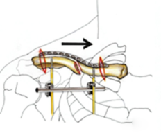
- Under fluoroscopic guidance, insert self-drilling Schanz screws, penetrating the cortex in both proximal and distal fragments. Care should be taken to avoid neurovascular damage during screw insertion.

- Once fracture reduction is confirmed, apply a connecting rod and tighten the nuts for temporary alignment.

- Make two small vertical incisions on the medial and lateral sides of the clavicle. During subcutaneous incisions and periosteal dissection, meticulous care should be taken to preserve neurovascular supply. A tunnel over the periosteum is established using a periosteal elevator along the superior surface of the clavicle.

- Pre-bend the plate and implant it through the periosteal tunnel.
- As stability is enhanced when the proximal fragment is connected to the axial skeleton, the first screw is applied at that location.

- The position of the distal fragment and the plate can be directly visualized through the lateral incision, ensuring proper plate placement on the bone. If necessary, reduction forceps can be used on the distal fragment followed by direct anterior plate fixation. After confirming satisfactory reduction, tighten the nuts. The second standard screw is fixed on the distal fragment.

- Remove the external fixator and tighten the distal screws.

- Use at least three screws to fixate the remaining screw holes in each bone fragment.
Case Presentation
1.
2.
Post time: Dec-27-2023