
Sports medicine guide | Knotless soft-tissue fixation
Knotless suture anchors are implants designed to secure suture or suture tape without requiring an arthroscopic knot. They can simplify selected soft-tissue-to-bone repair workflows and eliminate knot-stack-related concerns, but they are not universally superior to knotted anchors. Clinical suitability depends on the procedure, tissue pattern, bone quality, anchor material, fixation mechanism and complete repair construct.
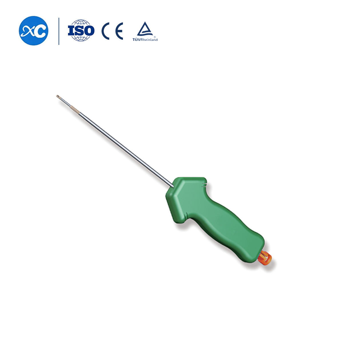
What Is a Knotless Suture Anchor?
A suture anchor is an implant used to connect soft tissue such as tendon, ligament or labrum to bone. In a conventional knotted repair, the surgeon places an anchor and ties arthroscopic knots to secure the tissue. A knotless anchor contains a mechanism that captures or locks the suture without requiring that final knot stack.
The term “knotless” does not mean that every anchor works in the same way. Some designs are push-in, others are threaded or screw-in, and some use an interference mechanism, internal locking element or separate tensioning step. The number of sutures or tapes, whether they are preloaded, and whether retensioning is possible depend on the exact system.
How the Knotless Mechanism Works
At a high level, the surgeon passes suture or tape through the target tissue, applies the planned tension and secures the construct within the anchor. The locking mechanism is intended to maintain the suture position after insertion. Anchor placement, tensioning sequence and whether the construct can be adjusted after insertion are product-specific.
Mechanical fixation supports healing; it does not create it. The anchor is intended to maintain soft-tissue position while biological integration develops. It cannot guarantee faster healing, pain relief or an earlier return to activity.
Knotless Does Not Describe the Material
A common source of confusion is treating “knotless,” “PEEK” and “all-suture” as interchangeable terms. They describe different characteristics:
- Knotless: a fixation mechanism that does not require a surgical knot.
- PEEK: a nonabsorbable thermoplastic polymer used for some rigid anchor bodies.
- Biocomposite: an absorbable or partially absorbable composite material used in selected anchor designs.
- All-suture: an anchor whose main fixation body is formed from flexible suture material.
- UHMWPE: a high-strength fiber commonly used in surgical suture or tape; it is not necessarily the anchor body.
Types of Knotless Suture Anchors
Knotless anchors are classified by material, insertion method, locking mechanism, size and intended anatomical application. Product families should be compared using their actual specifications rather than broad claims about the entire category.
PEEK Knotless Anchors
PEEK is a nonabsorbable polymer with radiolucent properties. Compared with metal, it generally produces less image artifact, although many anchor designs include radiopaque markers or other features that help assess position. Radiolucency does not prove that PEEK improves biological healing or lowers infection risk.
XC Medico's PEEK knotless suture anchors represent one material-and-mechanism combination. Current dimensions, suture configuration, instruments, intended use and regulatory status should be confirmed for the exact catalogue number.
Biocomposite and Absorbable Anchors
Biocomposite anchors are designed to change or resorb over time according to their material formulation. Potential considerations include degradation behavior, cyst formation, inflammatory response, imaging appearance and the biological response at the insertion site. These characteristics vary by material and product.
All-Suture Knotless Anchors
All-suture anchors expand or deploy within bone using a flexible suture-based body. Some designs require smaller drill holes than rigid anchors, which may be useful when bone preservation or multiple anchor placement is important. However, fixation still depends on appropriate deployment, cortical support or cancellous bone quality, insertion angle and the product-specific mechanism.
A flexible all-suture anchor should not be used to describe the structure of every PEEK knotless anchor. They are separate product categories with different insertion techniques and mechanical behavior.
| Anchor category | General characteristic | Important considerations |
|---|---|---|
| PEEK knotless | Rigid, nonabsorbable polymer body with a knotless locking mechanism | Anchor size, thread or push-in design, imaging markers, locking method and bone preparation |
| Biocomposite knotless | Rigid anchor made from an absorbable or composite material | Material degradation, biological response, insertion damage and long-term imaging appearance |
| All-suture knotless | Flexible suture-based fixation body that deploys within bone | Deployment, cortical or cancellous support, drill-hole size, pullout behavior and anchor expansion |
| Metal knotless | Nonabsorbable metallic anchor with a knotless suture mechanism | Imaging artifact, removal considerations, material sensitivity and insertion technique |
Common Surgical Applications
Knotless suture anchors are used for selected soft-tissue-to-bone repairs. The exact anatomical indications depend on the product's approved or registered labeling, so a shoulder anchor should not automatically be promoted for the knee, foot or ankle.
Rotator Cuff Repair
In arthroscopic rotator cuff repair, knotless anchors may be used in selected single-row, double-row or transosseous-equivalent constructs. The repair strategy depends on tear size and pattern, tendon mobility and quality, bone quality, footprint preparation and the planned distribution of suture or tape.
Knotless designs eliminate knot tying in the relevant part of the construct, but this does not prove that every knotless technique has greater initial strength or a lower retear rate. Laboratory behavior and clinical outcomes must be evaluated separately.
Bankart and Labral Repair
Knotless anchors can be used in selected capsulolabral repairs for shoulder instability. The surgeon may tension the tissue before locking the suture within the anchor. Outcomes are influenced by patient selection, bone loss, number and position of anchors, tissue quality, associated lesions and rehabilitation—not only whether the anchor is knotted or knotless.
Selected Tendon and Ligament Repairs
Depending on labeling, suture anchors may be used in selected biceps, elbow, hand, wrist, foot or ankle tendon and ligament repairs. The phrase “soft-tissue fixation” is broad, but each implant has a defined anatomical and procedural scope.
General knotless anchors should not automatically be described as primary ACL or PCL graft-fixation devices. Cruciate ligament reconstruction often uses specialized interference screws, cortical suspension devices, plates or other systems. Likewise, plastic surgery and general wound closure should not be added as indications without explicit product authorization.

Knotless vs Knotted Suture Anchors
Knotless and knotted repairs use different methods to secure suture, but neither is universally superior. Comparisons must involve the same procedure, repair configuration, tissue pattern and clinical endpoint.
Eliminates arthroscopic knot tying and knot stacks. Tension is controlled through the anchor-specific locking mechanism and insertion sequence.
Uses arthroscopic knots to secure the suture. It allows knot-based fixation but requires reliable knot technique and management of knot placement and bulk.
| Comparison factor | Knotless | Knotted |
|---|---|---|
| Suture security | Depends on the internal locking or interference mechanism | Depends on knot configuration, security and suture handling |
| Tension control | May allow controlled tensioning or retensioning, depending on design | Controlled during knot tying; tissue tension can change while knots are seated |
| Knot profile | No arthroscopic knot stack | Knot position and bulk require management |
| Workflow | May remove knot-tying steps | Requires arthroscopic knot-tying technique |
| Clinical outcomes | Current evidence in common shoulder procedures generally shows comparable outcomes rather than consistent superiority of one category | |
What the Clinical Evidence Shows
A 2025 systematic review and meta-analysis of arthroscopic Bankart repair found no significant differences between knotless and knotted techniques in redislocation, recurrent subluxation, revision surgery, anchor number or Rowe score improvement. Knotless repair may reduce operative time slightly, but the clinical significance remains uncertain.
For rotator cuff repair, meta-analyses also report broadly comparable pain, functional scores, retear rates and overall complications between knotted and knotless suture-bridge techniques. Some studies identify differences in retear pattern or time-zero biomechanics, which should not be converted into a universal clinical ranking.
Avoid misleading comparisons: a return-to-play or revision rate from one Bankart study cannot be used as a general result for every knotless anchor, rotator cuff repair, Achilles repair or ligament procedure.
Potential Advantages of a Knotless Technique
The most defensible advantages relate to surgical workflow and the absence of arthroscopic knots—not guaranteed patient outcomes.
These potential advantages do not automatically mean less pain, lower infection risk, finer scars, fewer revisions or faster biological healing. Those outcomes require procedure-specific clinical evidence.
General Knotless Anchor Repair Workflow
The following overview is educational and is not a surgical technique. Each implant must be used with its current instructions, compatible instruments and appropriate professional training.
Portal placement, insertion angle, bone preparation and tensioning cannot be standardized in a general blog. They vary by joint, repair pattern, anchor design and surgical technique. Knotless anchors also should not be described as automatically suitable for surgeons without arthroscopic training.
Risks and Limitations
Knotless fixation removes knot-related variables but introduces dependence on the anchor's locking mechanism and insertion technique. Potential issues include:
- Anchor pullout: fixation may fail if bone purchase, insertion or loading is inadequate.
- Suture slippage or locking failure: the intended tension may not be maintained if the mechanism is damaged or incorrectly deployed.
- Anchor breakage or insertion damage: excessive force, incorrect preparation or instrument mismatch may damage the implant.
- Malposition or prominence: incorrect trajectory or depth may affect fixation or nearby tissue and cartilage.
- Bone loss or cortical breach: repeated drilling, revision placement or an unsuitable anchor size can compromise the insertion site.
- Tissue cut-through: suture or tape can damage poor-quality tissue under load regardless of anchor security.
- Biological or material response: reactions, cyst formation or imaging changes may differ among PEEK, biocomposite and other materials.
- General surgical complications: infection, stiffness, pain, recurrent instability and failed healing remain possible.
Bone quality still matters. A self-locking suture mechanism does not make anchor fixation independent of bone. Pullout resistance depends on bone quality, anchor size and design, insertion site, trajectory, preparation and the loads applied by the repair.
How Do Surgeons Select a Knotless Anchor?
Selection should start with the procedure and tissue-repair objective. Relevant factors include:
- authorized anatomical indication and current product labeling;
- tissue type, tear pattern and tissue quality;
- bone density, available bone volume and previous anchor holes;
- anchor material, dimensions and insertion method;
- suture or tape material, width, number and configuration;
- locking mechanism and whether retensioning is supported;
- planned single-row, double-row, bridge or other repair construct;
- compatible guides, drills, punches and insertion instruments;
- revision considerations and postoperative imaging requirements;
- surgeon training and familiarity with the system.
FDA-cleared knotless PEEK anchor systems list specific soft-tissue-to-bone indications by anatomical region. This reinforces that “knotless” is not a blanket authorization for every arthroscopic, ACL, plastic-surgery or wound-repair procedure.
PEEK Knotless Anchors for Hospitals and Distributors
Professional buyers should evaluate the complete implant-and-instrument system rather than selecting an anchor solely because it is knotless or made from PEEK. XC Medico organizes available designs within its suture anchor product categories and broader orthopedic and sports medicine systems.
Related options such as non-absorbable suture anchors should be treated as separate fixation categories. For shoulder workflows, buyers may also review the compatible shoulder joint instrument set, while confirming compatibility with the exact anchor system.
| Procurement checkpoint | What to verify |
|---|---|
| Intended use | Exact anatomical indications and whether the configuration is authorized for arthroscopic or open repair |
| Anchor specification | Material, diameter, length, thread or push-in geometry and insertion depth |
| Suture configuration | Preloaded suture or tape, material, size, color, number of limbs and locking method |
| Instrumentation | Compatible guide, drill, punch, tap, inserter and replacement instrument availability |
| Testing | Applicable pullout, cyclic displacement, locking and insertion testing for the final configuration |
| Documentation | Instructions for use, sterile status, shelf life, certificates, traceability and market registration |
For professional procurement: request the current PEEK knotless anchor catalogue, implant specifications, compatible instrument list and destination-market documentation before choosing a configuration.
Frequently Asked Questions About Knotless Suture Anchors
What is the main difference between knotless and knotted suture anchors?
A knotless anchor secures the suture through an internal locking or interference mechanism. A knotted repair uses arthroscopic knots to secure the suture after anchor placement.
Are knotless anchors better than knotted anchors?
Not universally. Current evidence in Bankart and rotator cuff repairs generally shows comparable clinical outcomes. Knotless systems may simplify workflow or reduce knot bulk, while each approach has technique-specific considerations.
Are all knotless anchors made from PEEK?
No. Knotless anchors may be made from PEEK, biocomposite materials, metal or flexible all-suture constructs. “Knotless” describes how the suture is secured, not the anchor material.
Does a PEEK anchor improve healing?
PEEK offers material and imaging characteristics, but it does not independently guarantee faster biological healing. Healing depends on the tissue, fixation construct, bone quality, rehabilitation and patient factors.
Can knotless anchors be used in every arthroscopic procedure?
No. Use depends on the exact anchor's labeled anatomical indications, size, material, compatible instruments and the planned repair technique.
What should distributors verify before sourcing knotless anchors?
Verify intended use, anchor and suture specifications, instruments, mechanical testing, sterile status, traceability, instructions for use and regulatory status for the destination market.
Medical and Regulatory References
- No Clear Difference in Clinical Outcomes Between Knotted and Knotless Arthroscopic Bankart Repair: A Systematic Review.
- Knotless versus Knotted Arthroscopic Bankart Repairs: A Systematic Review and Meta-Analysis.
- Clinical Outcomes of Knotted versus Knotless Suture-Bridge Rotator Cuff Repair: A Meta-Analysis.
- PEEK Knotless and Knotted Biodegradable Suture Anchors in Anterior Shoulder Instability: A Randomized Study.
- U.S. FDA 510(k) Documentation: Knotless PEEK Suture Anchor Intended Uses.

