


Orthopedic Sports Medicine Guide
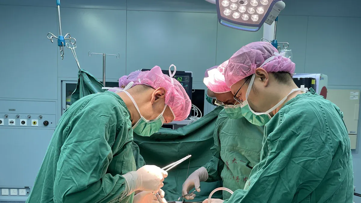
Suture anchors are small implants used to attach soft tissue—such as a tendon, ligament, or labrum—to bone. They are commonly used in open and arthroscopic orthopedic surgery, including rotator cuff repair, shoulder stabilization, and selected knee, foot, ankle, hand, and elbow procedures.
Quick answer: A surgeon places the anchor in bone and uses the attached suture to hold the injured soft tissue against the bone while healing occurs. Whether a suture anchor dissolves depends on its material. Titanium, stainless-steel, and PEEK anchors are non-absorbable, while bioabsorbable and some biocomposite anchors are designed to degrade gradually. Degradation can take months or years and may not be complete in every case.
Key Takeaways
What Is a Suture Anchor?
In orthopedic surgery, an “anchor” is a fixation device inserted into bone to provide an attachment point for one or more sutures. Those sutures pass through or around the damaged soft tissue and hold it against bone. The objective is to maintain the repair position while biological healing develops; the implant itself does not heal the tissue.
A typical suture anchor system includes:
The anchor body: the component fixed in bone. Depending on its design, it may be threaded, press-fit, expanding, or made primarily from suture material.
The eyelet or suture-retention mechanism: the part that connects the anchor and suture. In some knotless designs, the mechanism also tensions and locks the repair.
The suture or tape: the material that passes through the tissue. Wider tape-type sutures may be selected for some repair constructs, although biomechanical differences do not automatically guarantee better clinical outcomes.
How Do Suture Anchors Work?
The exact technique varies by operation and anchor design, but the basic fixation process follows the same principles:
The surgeon prepares an insertion point in the bone near the intended tissue attachment site.
The anchor is inserted at the planned position and angle using the appropriate delivery instrument.
The suture is passed through the tendon, ligament, capsule, or labrum.
The suture is tied or secured with a knotless locking mechanism to bring the tissue into contact with bone.
The repair protects the tissue-to-bone interface during the early healing period, while postoperative rehabilitation controls load and motion.
Fixation strength is influenced by more than the anchor alone. Bone quality, insertion angle, anchor diameter and design, suture configuration, tissue condition, surgical technique, and rehabilitation all affect the repair. An anchor that is appropriate for one anatomical site or patient may not be appropriate for another.
Important: Suture anchors can support minimally invasive techniques, but they do not by themselves guarantee less pain, faster healing, or a specific clinical result. Recovery depends on the original injury, procedure, patient health, repair quality, and rehabilitation plan.
Do Suture Anchors Dissolve?
Some suture anchors are designed to degrade, while others remain in the body. “Dissolvable” is a useful everyday description, but bioabsorbable or biodegradable is more precise. These anchors break down through biological and chemical processes rather than disappearing immediately.
| Anchor material or design | Does it dissolve? | Practical consideration |
|---|---|---|
| Metal, such as titanium or stainless steel | No | Provides permanent radiopaque fixation but may create imaging artifact and can complicate revision in some situations. |
| PEEK | No | A non-absorbable polymer that is radiolucent on standard X-rays and generally creates less imaging artifact than metal. |
| Bioabsorbable polymer | Designed to degrade | The rate varies substantially by polymer composition, anchor size, location, and biological response. |
| Biocomposite | Often designed to degrade gradually | Combines a degradable polymer with a mineral component; replacement by bone is variable and should not be assumed. |
| All-suture anchor | Depends on the material | “All-suture” describes construction, not necessarily absorption. Product-specific material information must be checked. |
Bioabsorbable anchors do not all degrade on the same schedule. Depending on the material, remnants may be visible for years, and complete absorption is not guaranteed. Anchor selection should therefore be based on the clinical situation and product documentation—not simply on a preference for an implant that “dissolves.”
Types of Suture Anchor Devices

Suture anchor designs vary in material, geometry, insertion method, and suture-retention mechanism.
Non-Absorbable Anchors
Non-absorbable anchors are intended to remain in the body unless removal becomes medically necessary. The category includes metal designs as well as non-absorbable polymer designs such as PEEK. Non-absorbable anchors may provide long-term fixation, but the consequences for imaging and possible revision depend on the material and placement.
| Type of Suture Anchor | Potential advantages | Points to consider |
|---|---|---|
| Non-absorbable | Permanent material; available in multiple established designs and materials. | Material remains in the body; imaging artifact and revision considerations vary. |
| Bioabsorbable or biocomposite | Designed for gradual degradation; some designs may reduce permanent implant material. | Degradation is variable; inflammatory response, cyst formation, or bone changes can occur. |
| All-suture | Uses a small bone tunnel and may preserve more bone than some solid anchors. | Performance depends on deployment, bone quality, and design; absorption cannot be inferred from the name. |
PEEK Corded Anchors
PEEK corded suture anchor devices use polyether ether ketone, a non-absorbable engineering polymer. PEEK is radiolucent on standard radiographs and generally creates less imaging artifact than metal, although the implant position may be identified by associated markers or the surrounding bone tunnel. PEEK anchors do not dissolve.
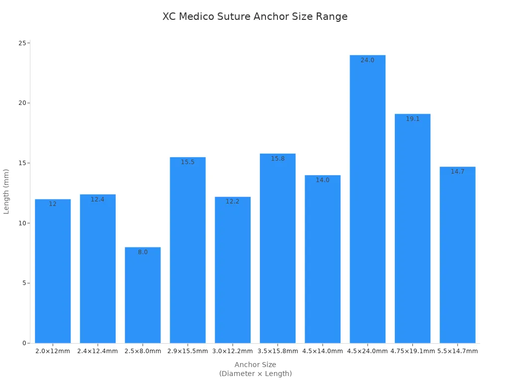
Anchor size is only one selection factor; anatomy, bone quality, tissue, and the planned repair construct must also be considered.
Knotless Anchors
Knotless anchors use a locking mechanism to secure and tension the suture without a conventional arthroscopic knot. Knot-free anchors can simplify certain repair steps and avoid knot-related prominence, but their suitability depends on the procedure and the surgeon’s fixation strategy. “Knotless” describes how the suture is secured; it does not identify whether the anchor is metal, PEEK, absorbable, or biocomposite.
All-Suture Anchors
All-suture anchors are formed mainly from high-strength suture material rather than a rigid anchor body. They typically require a smaller bone tunnel and expand or deploy beneath the cortical surface. Their behavior depends on deployment technique and local bone quality. They should not automatically be described as dissolvable.
Suture anchors are available in different sizes, materials, and loading configurations. XC Medico supplies orthopedic sports-medicine products, including selected PEEK and titanium-alloy suture anchor options. Product choice should be based on the intended use, local regulatory status, compatible instruments, and the responsible clinician’s judgment.
Where Are Suture Anchors Used?
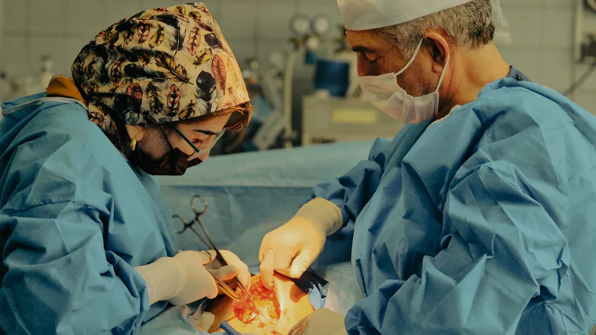
Suture anchors may be incorporated into procedures involving:
Shoulder: rotator cuff repair, Bankart or labral repair, and selected biceps procedures.
Hip: selected labral repairs.
Knee: selected ligament, tendon, meniscal-root, or capsular fixation procedures.
Foot and ankle: selected Achilles tendon, ligament, and tendon repairs.
Elbow, wrist, and hand: selected tendon or ligament reattachment procedures.
The surgeon decides whether an anchor-based repair, a transosseous technique, or another method is most appropriate. Clinical outcome is influenced by the injury pattern and overall repair—not merely by the presence of a particular anchor material.
Safety, Longevity, and Possible Complications
Suture anchors are widely used, but no implant or surgical procedure is risk-free. A properly selected and positioned anchor is intended to remain stable during healing. Non-absorbable anchors may remain permanently, whereas absorbable materials change over time at material-specific rates.
Potential complications can include:
Anchor pullout, loosening, migration, breakage, or loss of fixation
Suture abrasion, breakage, or knot-related irritation
Cartilage damage if an anchor is prominent or enters the joint
Bone reaction, tunnel enlargement, cyst formation, or osteolysis
Inflammatory or foreign-body response to certain materials
Infection, stiffness, persistent pain, or repair failure
Risk varies with the procedure, implant material, surgical technique, tissue quality, bone quality, rehabilitation, and patient factors. New or worsening pain, redness, drainage, fever, swelling, mechanical catching, or unexpected loss of motion after surgery warrants prompt assessment by the treating clinical team.
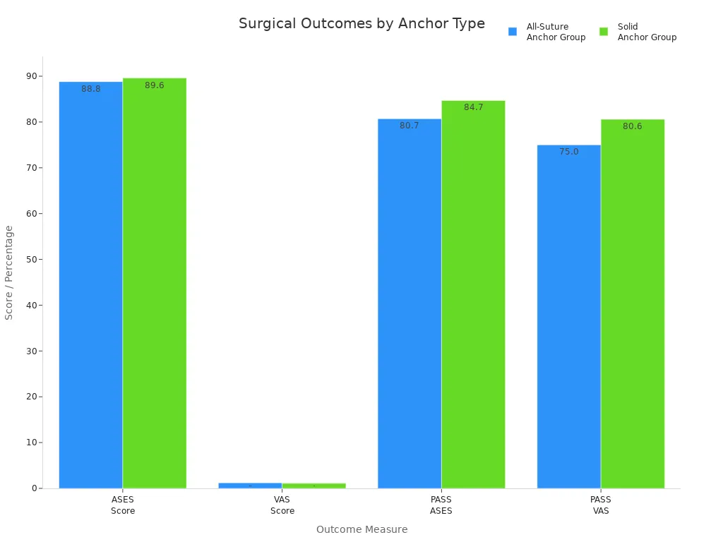
Clinical study results should be interpreted in the context of the procedure, patient population, follow-up period, and study design.
How Surgeons Evaluate an Anchor
When choosing a fixation system, the surgical team may consider:
The anatomical site and available bone volume
Cortical and cancellous bone quality
The amount and condition of tissue being repaired
Anchor diameter, length, geometry, and insertion method
Metal, PEEK, bioabsorbable, biocomposite, or all-suture construction
Knotted or knotless repair technique
Suture or tape size, configuration, and compatibility
Revision strategy and postoperative imaging needs
Regulatory clearance, traceability, sterile packaging, and instrument compatibility
Product development continues to refine materials, insertion systems, knotless mechanisms, and bone-preserving designs. However, “newer” does not automatically mean clinically superior. Claims about faster surgery, lower complication rates, stronger healing, or improved outcomes should be supported by product-specific biomechanical or clinical evidence.
Manufacturers should provide clear technical documentation and consistent quality controls. XC Medico supports orthopedic product development and new technology in suture anchors, but healthcare professionals and distributors should still verify the specifications, evidence, intended use, and registration status of each product in their market.
Suture Anchor FAQ
What does “anchor” mean in orthopedic surgery?
An anchor is a small implant fixed in bone to hold sutures. The sutures secure a tendon, ligament, capsule, or labrum against bone while the repaired tissue heals.
Do suture anchors dissolve?
Some do and some do not. Titanium, stainless-steel, and PEEK anchors are non-absorbable. Bioabsorbable and some biocomposite anchors are designed to degrade gradually, but this can take months or years and may not be complete in every patient.
Is an anchor stitch the same as a suture anchor?
No. “Anchor stitch” generally refers to a stitch pattern or suturing technique. A suture anchor is an implant inserted into bone to provide a fixation point for the suture.
How long do suture anchors stay in the body?
Non-absorbable anchors may remain permanently unless removal becomes medically necessary. Absorbable anchors degrade at different rates according to their material, location, and the patient’s biological response.
Can you feel a suture anchor?
Most anchors are seated within bone and are not directly felt. Postoperative discomfort can still arise from healing tissue, stiffness, inflammation, prominence, implant complications, or other causes. Persistent or worsening symptoms should be reviewed by the treating clinician.
Can suture anchors be seen on X-rays or MRI?
Metal anchors are usually visible on X-rays and may create imaging artifact. PEEK is radiolucent and is generally less visible on standard X-rays. Visibility and MRI artifact vary by material and product design, so patients should tell imaging staff about any implanted device.
What should you expect after suture anchor surgery?
Recovery varies by the repaired tissue and procedure. Patients may need temporary protection, restricted motion, and progressive rehabilitation. Return to work, exercise, or sport should follow the surgeon’s and rehabilitation team’s instructions rather than a fixed timetable.
When should you contact your doctor after surgery?
Contact the treating team for increasing pain, redness, drainage, fever, unusual swelling, mechanical catching, sudden weakness, or an unexpected loss of motion. Urgent symptoms should be assessed promptly.
Discuss Your Suture Anchor Requirements
For product specifications, material options, compatible instrumentation, or OEM manufacturing discussions, XC Medico gives safe and trusted products for you.
Medical disclaimer: This article is for general educational and product-information purposes. It does not provide diagnosis, treatment advice, or a recommendation for a specific implant. Patients should consult a qualified healthcare professional. Product availability, intended use, and regulatory status vary by market.

