


Orthopedic trauma guide | External fixation indications and frame types
Orthopedic surgeons use external fixation devices for selected open fractures, severe soft-tissue injuries, rapid temporary stabilization, some fractures near joints, limb reconstruction, deformity correction, limb lengthening and selected fusion procedures. Depending on the injury and treatment plan, the frame may be a temporary bridge to internal fixation or the definitive method used while the bone heals.
What Is an External Fixation Device?
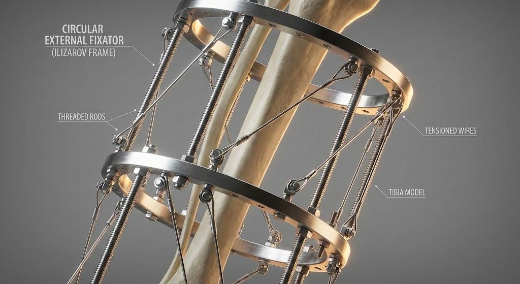
An external fixation device is an orthopedic frame used to stabilize or gradually correct bone position. Pins or wires are placed into bone and connected to bars, rings, clamps or other components outside the body. Because the main frame remains external, surgeons can access soft tissues and, with selected systems, adjust alignment during treatment.
External fixation describes a treatment method rather than one universal product. Frames range from compact devices for the hand or wrist to monolateral bars and circular systems used in lower-limb trauma and reconstruction. Device design, pin configuration and postoperative instructions must match the specific anatomy, intended use and authorized system.
Important distinction: an external fixator is not automatically a temporary device. It may be used temporarily in staged trauma care, or it may serve as the definitive fixation method until sufficient healing or correction is achieved.
Which Orthopedic Surgeries Use External Fixators?
The clearest answer is that external fixation is used when surgeons need skeletal stability while limiting additional disruption around an injured or reconstructed area. It is particularly useful when access to wounds or soft tissues must be preserved, when rapid provisional stabilization is needed, or when gradual correction is part of the treatment plan.
Severe Open Fractures and Soft-Tissue Injury
Open fractures communicate with a wound and may involve contamination, tissue damage and infection risk. External fixation can provide stability while surgeons perform debridement, wound management and soft-tissue reconstruction. In staged care, the initial frame may later be replaced with another fixation method after the tissues and patient are ready.
External fixation may also be considered when swelling, skin injury or compromised soft tissue makes immediate internal fixation undesirable. It does not independently prevent infection or guarantee healing; appropriate debridement, antibiotics when indicated, wound care, stable fixation and follow-up all contribute to treatment.
Temporary Damage-Control Orthopedics
In patients with multiple injuries or physiologic instability, a rapidly applied external frame can temporarily stabilize a long-bone or pelvic injury while the trauma team addresses life-threatening problems. This approach is commonly called damage-control orthopedics.
The decision to convert to internal fixation, revise the frame or retain external fixation depends on the patient's recovery, contamination, soft-tissue condition, alignment and overall surgical strategy. There is no universal conversion date that applies to every injury.
Tibial Plateau, Pilon and Other Periarticular Fractures
High-energy fractures near the knee or ankle can be accompanied by substantial swelling and soft-tissue injury. A joint-spanning frame may maintain length and alignment while swelling improves and definitive planning continues. In selected cases, a circular, hybrid or other external system may form part of the definitive reconstruction.
Because a spanning frame may restrict joint movement, pin position, frame geometry, soft-tissue access and the next stage of treatment must be considered before application.
Limb Lengthening and Deformity Correction
Circular and specialized external frames can be used after a planned bone cut to gradually change length, angulation, rotation or translation. Correction schedules are prescribed by the treating limb-reconstruction team and are adjusted according to radiographs, bone formation, soft-tissue tolerance, nerve function and joint motion.
A commonly discussed distraction rate must not be treated as a universal patient instruction. The appropriate rate and rhythm vary with anatomy, age, correction goal and biological response.
Joint Fusion, Nonunion and Limb Reconstruction
External fixation may be used in selected arthrodesis procedures, infected or complex nonunion treatment, bone transport and reconstruction after bone loss. These are specialist indications that require careful planning of stability, compression or distraction, bone biology, infection management and rehabilitation.
Mini external fixation systems may also be used for selected fractures, dislocations or reconstructive procedures involving the hand, wrist, foot or ankle when the product's intended use and the surgical plan support that application.
| Clinical situation | Possible role of external fixation | Common planning considerations |
|---|---|---|
| Open fracture with soft-tissue damage | Temporary stabilization or selected definitive fixation | Wound access, contamination, alignment and soft-tissue coverage |
| Polytrauma or unstable patient | Rapid damage-control stabilization | Physiologic condition, later conversion strategy and frame access |
| High-energy periarticular fracture | Joint-spanning temporary frame or selected definitive construct | Swelling, joint surface injury, pin placement and future incisions |
| Deformity correction or limb lengthening | Gradual postoperative correction | Correction plan, regenerate bone, nerves, muscles and joint motion |
| Nonunion, infection or bone loss | Stability, compression, bone transport or staged reconstruction | Bone biology, infection control, defect size and patient tolerance |
| Selected joint fusion | Compression and maintenance of alignment | Bone contact, frame stability, soft tissue and weight-bearing plan |
Temporary vs Definitive External Fixation
Used to obtain early stability while the patient, wound or soft tissues recover. The treatment plan may later change to internal fixation or another reconstructive method.
The frame remains the principal fixation construct during fracture healing, fusion or gradual correction and is removed when the treating team determines that the treatment objective has been met.
Temporary does not mean unimportant, and definitive does not mean permanent. Both strategies require accurate pin placement, sufficient frame stability, appropriate alignment and a documented follow-up plan.
AAOS notes that an external fixator may be applied when skin and soft tissues are badly damaged until the patient can tolerate surgery, while in other cases it may be the definitive treatment. The correct pathway is therefore case-specific rather than determined by the device name alone.
Types of External Fixation Frames
Frame type is selected according to the anatomical region, fracture or reconstruction pattern, required stability, need for adjustment and product-specific intended use. XC Medico groups its available products within its orthopedic external fixation systems.
Monolateral External Fixators
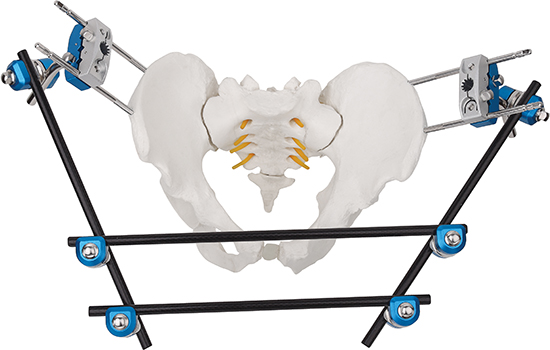
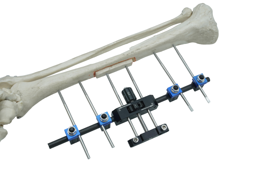
A monolateral frame is positioned mainly along one side of the limb. Depending on the system, it may offer a comparatively straightforward bar-and-clamp configuration and access around much of the limb. It can be used in selected trauma and reconstructive applications when sufficient stability can be achieved.
Professional buyers can review the available monolateral external fixation systems, but the exact indication and configuration must be confirmed from current labeling and surgical technique documents.
Circular and Ilizarov Frames
Circular frames use rings connected to tensioned wires, half pins and rods or struts. Their geometry can support multiplanar stability and gradual correction, making them relevant to selected complex fractures, deformity correction, bone transport and limb reconstruction.
Ilizarov circular external fixators are one family within this category. Assembly and correction protocols require specialist training and should not be inferred from a general product photograph.
Modular and Hybrid Fixators
Modular systems allow components to be arranged for different fracture locations and access needs. Hybrid constructs may combine rings, wires, half pins or unilateral components to meet a selected stability and anatomy requirement.
Mini and Joint-Spanning Fixators
Mini frames can be configured for selected small-bone injuries, while joint-spanning constructs can temporarily bridge a severely injured knee, ankle, wrist or other joint. A spanning frame may help maintain alignment but can restrict joint motion, so the duration and rehabilitation plan require close supervision.
Bone quality matters: external fixation pins and wires must obtain adequate purchase in bone. Osteoporosis does not automatically make external fixation preferable; poor bone quality can complicate fixation and must be considered in device selection, pin strategy and the overall treatment plan.
How Is an External Fixator Applied?
External fixation is performed in an operating-room setting using an appropriate anesthesia and imaging plan. The exact steps differ by body region, frame design and whether the goal is temporary stabilization, definitive fixation or gradual correction.
Pin placement must avoid important nerves, vessels, joints and damaged soft tissues. When another operation is anticipated, pin sites should also be planned so they do not interfere with later incisions or implants. These details are determined by the surgical team and cannot be replaced by general online instructions.
How Do Surgeons Select an External Fixation System?
There is no single “best” external fixator for every orthopedic surgery. Selection should follow the indication, anatomy and required mechanical behavior of the complete construct.
Product pages such as the ankle joint external fixator can help professional users identify a product family. They do not replace the applicable labeling, surgical technique, local registration or patient-specific clinical judgment.
Recovery, Weight Bearing and Frame Duration
Recovery varies widely. Frame duration may be influenced by fracture pattern, bone healing, soft-tissue reconstruction, infection, correction goal, age, health and the mechanical environment. A fixed number of weeks or months should not be promised for all patients.
Weight-bearing instructions are also individualized. Some constructs are designed to permit selected loading, while others require partial or non-weight bearing. Patients should follow the written instructions of their orthopedic and rehabilitation teams rather than assuming that the presence of a frame makes walking safe.
Follow-up commonly considers:
- radiographic alignment and evidence of healing or regenerate bone formation;
- frame and pin stability;
- pain, swelling, skin condition and pin-site appearance;
- nerve and blood-vessel function;
- movement of nearby joints and muscle function;
- progress toward the prescribed loading and rehabilitation goals.
After frame removal, some patients require protection, bracing or progressive rehabilitation. Timing and activity progression depend on the treating team's assessment of bone stability and function.
Risks, Complications and Pin-Site Care
External fixation can provide important treatment advantages in selected cases, but it also has risks. Pin-site irritation or infection is among the most common concerns. Other possible complications include pin loosening, loss of alignment, delayed union, nonunion, malunion, joint stiffness, muscle or tendon tethering, nerve or blood-vessel injury, hardware failure and deep infection.
Pin-care protocols differ among hospitals and frame teams, and evidence does not support one universal cleaning method for every situation. Patients should use the supplies, frequency and technique taught by their own clinical team.
Contact the treating team promptly for increasing pain around a pin, spreading redness, increasing swelling, pus or unusual discharge, fever, feeling unwell, new difficulty bearing weight, a loose component, a change in limb position, new numbness or a pale or cold limb. Severe symptoms or suspected neurovascular compromise require urgent medical assessment.
Frames should not be adjusted, tightened or removed by the patient unless the treating team has provided a specific prescribed adjustment schedule and training. Any unexpected frame movement or damaged component should be reported.
External Fixation Systems for Hospitals and Distributors
For hospitals, distributors and OEM/ODM partners, evaluation should cover the complete system rather than a single bar, ring or clamp. Procurement decisions should verify market-specific registration, intended use, material documentation, traceability, mechanical testing, labeling, cleaning or sterilization instructions, compatible pins and instruments, packaging configuration and after-sales support.
XC Medico's external fixation device categories include modular, monolateral, circular and anatomy-specific options. Availability and regulatory status may differ by market, so buyers should request current documentation for the exact catalogue number and destination country.
| Procurement checkpoint | Questions to verify |
|---|---|
| Intended use | Which bones, procedures and frame configurations are included in the current product labeling? |
| System completeness | Are compatible pins, wires, clamps, rods, rings, instruments and replacement components available? |
| Documentation | Can the supplier provide certificates, testing records, instructions for use and lot-level traceability? |
| Sterile status | Which components are supplied sterile or non-sterile, and what validated processing instructions apply? |
| Training and support | Are assembly guidance, instrument lists, product training and complaint-handling processes available? |
| Market access | Is the exact device configuration authorized for the destination market and proposed indication? |
For professional procurement: review the available external fixator families and request the current catalogue, component list and market-specific regulatory documents before selecting a system.
Frequently Asked Questions About External Fixation
What orthopedic surgeries commonly use external fixation?
External fixation may be used for selected open fractures, severe soft-tissue injuries, damage-control stabilization, high-energy periarticular fractures, limb lengthening, deformity correction, nonunion or bone-loss reconstruction, and selected joint-fusion procedures.
Is an external fixator always temporary?
No. Some frames are temporary bridges to another operation, while others are intended to remain as the definitive fixation construct during healing or correction.
Can a patient walk with an external fixator?
Only when the treating surgeon and rehabilitation team permit it. Weight-bearing status depends on the injury, frame configuration, bone quality, healing and overall treatment plan.
How long does an external fixator stay on?
There is no universal duration. The frame remains according to the objective of treatment, radiographic progress, soft-tissue condition, stability and the treating team's assessment.
What is the most common concern during external fixation?
Pin-site irritation or infection is a common concern. Patients need individualized pin-care instructions and should report increasing pain, spreading redness, swelling, pus, fever or frame loosening promptly.
Is external fixation safer than internal fixation?
Neither method is universally safer. Each has different indications, benefits and risks. The appropriate choice depends on the fracture, soft tissues, contamination, patient condition and reconstructive plan.
Medical and Regulatory References
- American Academy of Orthopaedic Surgeons: Fractures (Broken Bones).
- U.S. Food and Drug Administration: Reviewer's Guidance for Orthopedic External Fixation Devices.
- Royal National Orthopaedic Hospital: External Fixation for Bone Correction and Lengthening.
- Royal National Orthopaedic Hospital: Pin-Site Care Guide.

