K-Wire Fixation for Distal Radius Fractures: Principles, Indications and Risks
Views: 61 Author: Site Editor Publish Time: 2026-08-20 Origin: Site
Percutaneous K-wire fixation is one of several methods used to maintain reduction in selected distal radius fractures. Also called closed reduction and percutaneous pinning, the technique uses Kirschner wires to stabilize fracture fragments after alignment has been restored.
It is not a universal solution for every broken wrist. Fracture pattern, reduction stability, articular involvement, bone quality, soft-tissue condition, patient priorities and available fixation options all influence treatment selection. This guide explains the principles and limitations of distal radius K-wire fixation without replacing formal surgical training or a device-specific technique guide.
Key Takeaways
K-wire fixation is generally considered after fracture reduction when additional stabilization is required and the fragments can be controlled with wires.
Radial styloid, dorsal, fragment-specific and intrafocal configurations describe different principles rather than one mandatory pin pattern.
Wire number, diameter, trajectory and immobilization strategy are case- and device-specific; they should not be selected from a general marketing article.
Potential concerns include loss of reduction, pin-tract infection, wire migration, soft-tissue irritation, tendon or nerve injury and wrist stiffness.
K-wires, volar plates and external fixation each have different tradeoffs. Current evidence does not support one fixation method as universally superior for every unstable or complete articular fracture.
What Is K-Wire Fixation for a Distal Radius Fracture?
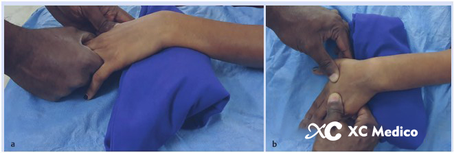
A distal radius fracture occurs near the wrist end of the radius. When a fracture is displaced, clinicians assess whether acceptable alignment can be restored and maintained. Closed reduction refers to restoring alignment without opening the fracture site. Percutaneous pinning then places one or more smooth wires through small skin entry points to hold selected fragments while healing progresses.
The preferred medical term is closed reduction and percutaneous pinning, not “closed needle insertion.” A Kirschner wire is a fixation device rather than an injection needle. The article should therefore use K-wire, wire, pinning and fixation terminology consistently.
How K-Wires Maintain Reduction
Wires may cross a fracture and gain purchase in a stable portion of the radius, or they may support individual fragments in a configuration chosen for the fracture pattern. Mechanical stability depends on more than the number of wires. Divergence, fragment control, cortical purchase, wire diameter, bone quality and supplemental immobilization can all affect the construct.
K-Wire Fixation Is One Treatment Option
Some distal radius fractures can be managed without surgery, while others may be treated with percutaneous pins, a volar or dorsal plate, an external fixator or a combined strategy. For a wider overview, see XC Medico’s guide to the diagnosis, classification and treatment of distal radius fractures.
Where Percutaneous Pinning Fits in Treatment
Nonoperative Management
Selected stable fractures or fractures that remain acceptably aligned after reduction may be managed with a cast or splint and follow-up imaging. Patient age alone does not determine treatment.
Percutaneous K-Wire Fixation
Pinning can provide additional fragment control after reduction in selected patterns. It usually requires attention to pin sites, immobilization and later wire management.
Plate Fixation
A volar or dorsal plate provides internal fixation and may be selected for patterns requiring direct fragment control. XC Medico supplies several distal radius locking plate options.
External Fixation
A spanning or nonspanning frame may help maintain length and alignment in selected unstable fractures, including cases in which soft-tissue conditions affect internal fixation planning.
The AAOS/ASSH clinical practice guideline reports no significant difference in radiographic or patient-reported outcomes among fixation techniques for complete articular or unstable distal radius fractures, although volar locked plates may provide earlier functional recovery in the short term. This supports individualized selection rather than a claim that K-wires or plates are always better.
When May Percutaneous K-Wire Fixation Be Considered?
Clinical decision-making begins with examination and imaging. The following factors may be considered by qualified professionals, but they are not a stand-alone indication checklist.
Fracture Pattern and Reduction Stability
Pinning is more likely to be useful when key fragments can be reduced and controlled by appropriately positioned wires. Marked metaphyseal comminution, small articular fragments or a tendency to collapse may reduce the reliability of a simple wire construct and prompt consideration of another or supplemental method.
Bone Quality and Fragment Size
Osteoporotic bone can provide less reliable wire purchase. Adding wires is not automatically sufficient to solve poor fixation, and there is no universal rule that every osteoporotic fracture requires four or five pins. Construct selection should follow the fracture pattern, imaging, approved technique and surgeon judgment.
Patient and Soft-Tissue Factors
Skin condition, swelling, open injury, medical comorbidities, activity demands, ability to follow pin-site and immobilization instructions and expected rehabilitation all contribute to treatment selection.
Age and Functional Demand
Evidence should be interpreted alongside patient-level factors. The AAOS/ASSH guideline reports that operative treatment in geriatric populations does not generally improve long-term patient-reported outcomes compared with nonoperative care, even when radiographic alignment differs. Shared decision-making remains important.
Clinical boundary: a general educational article cannot determine whether a specific fracture requires K-wires, a plate, external fixation or nonoperative treatment. That decision requires patient assessment, imaging and qualified medical judgment.
Common K-Wire Fixation Principles
Several pinning concepts are described in orthopedic practice. The names below identify broad strategies; they should not be interpreted as fixed entry points, angles or mandatory wire counts.
Radial Styloid Pinning
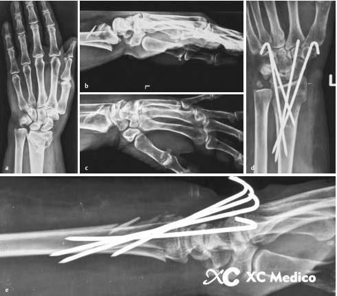
A radial styloid wire may be directed across the fracture to gain purchase in the proximal radius. The sensory branch of the radial nerve, tendons, joint surface and wire position require consideration. Exact trajectory depends on anatomy, fracture morphology and the selected technique.
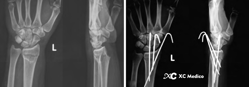
Illustrative distal radius pinning configuration. Final wire placement must be based on fracture anatomy, fluoroscopic assessment and the approved surgical technique.
Dorsal and Fragment-Specific Pinning
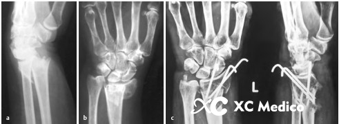
Dorsal wires or wires directed toward a lunate facet fragment may be used when those fragments require control. Dorsal anatomy includes extensor compartments and tendons that can be irritated or injured by an unsuitable entry point, prominent wire or migration.
Example of fragment-oriented wire fixation. The illustration describes a principle, not a universal three-wire template.
Kapandji Intrafocal Pinning
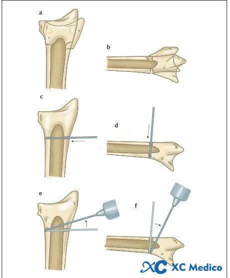
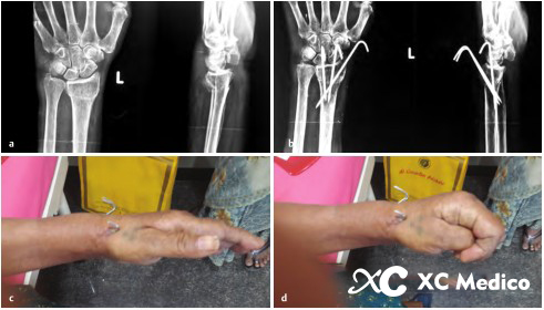
In the Kapandji technique, wires are introduced at the fracture site and used to assist reduction and support the distal fragment before being advanced to maintain the construct. The method is distinct from simply crossing the fracture from a distant entry point. Suitability depends on the fracture configuration and bone quality.
Kapandji intrafocal pinning concept. Specific wire size, number and direction should come from a validated technique and case-specific planning.
Supplemental Pinning and Combined Fixation
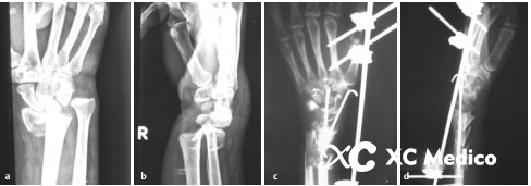
Complex fractures may require additional fragment-specific control or a combined construct. However, more wires do not automatically produce better stability. Wire convergence, insufficient spread, poor cortical purchase or inability to control a key fragment can still result in loss of reduction.
Comminution can make simple pinning less reliable. Supplemental or alternative fixation may be evaluated according to imaging and patient factors.
Imaging and Fixation Assessment
Reduction and Articular Alignment
Imaging is used to evaluate radial height, inclination, tilt, articular congruity and distal radioulnar joint relationships. The acceptable target depends on the patient and fracture. A technically placed wire does not compensate for an unacceptable reduction.
Wire Position and Cortical Purchase
Fluoroscopic views help assess whether wires control the intended fragments, avoid the radiocarpal and distal radioulnar joint spaces where required, and obtain suitable purchase without problematic prominence. Multiple views may be needed because a wire that appears acceptable in one projection may not be acceptable in another.
Construct Stability
Stability assessment considers the fracture, wire configuration and planned immobilization together. Wires clustered in one region may provide less rotational control than a well-distributed construct, but a generalized spacing rule should not replace biomechanical and clinical judgment.
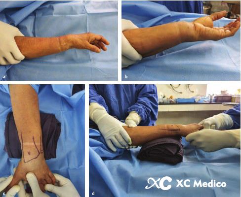
Wire trajectory and fracture control should be assessed in appropriate imaging views before the construct is accepted.
Potential Complications and Risk Considerations
Complications cannot be completely prevented by a checklist. The goal is to recognize relevant risks and follow the specific implant instructions, sterile protocol and postoperative plan.
Potential concerns include:
Loss of reduction or fixation failure
Pin-tract infection or deeper infection
Wire loosening, migration, breakage or skin irritation
Injury or irritation involving extensor tendons, sensory nerves or other soft tissues
Joint penetration or unsuitable cortical prominence
Wrist and finger stiffness
Malunion, post-traumatic arthritis or complex regional pain syndrome
Loss of Reduction
Comminution, poor bone quality, insufficient fragment control, unsuitable wire configuration or inadequate immobilization may contribute to secondary displacement. Follow-up imaging may be used according to the treating team’s protocol and the stability of the fracture.
Pin-Tract Infection
Pin-site condition should be monitored according to institutional and surgeon instructions. Increasing redness, drainage, swelling, pain, fever or other concerning symptoms require clinical evaluation. This article does not prescribe a universal pin-care regimen.
Tendon, Nerve and Soft-Tissue Injury
Entry location, wire trajectory, prominence and migration can place nearby structures at risk. Lister’s tubercle and the dorsal extensor compartments are clinically relevant landmarks, while the radial sensory nerve requires consideration near radial-sided entry points.
Thermal and Mechanical Tissue Injury
Repeated drilling, excessive heat, unsuitable equipment or poor technique may damage bone or soft tissue. Qualified users should follow the approved technique, equipment instructions and institutional protocols rather than generic online operating steps.
Post-Fixation Follow-Up Considerations
Follow-up varies with fracture stability, fixation method and patient factors. Common areas assessed include wound or pin-site condition, neurovascular status, finger motion, swelling, pain, radiographic alignment and signs of wire loosening or migration.
Immobilization duration, rehabilitation, wire removal and return to activity are individualized. Patients should not change a splint, perform exercises or manage an exposed wire based solely on online information.
Finger motion and soft-tissue condition may be reviewed during follow-up, while wrist rehabilitation depends on construct stability and the treating clinician’s plan.
K-Wires vs Volar Plates vs External Fixation
Method
General Characteristics
Important Considerations
Percutaneous K-wire fixation
Uses relatively small percutaneous implants to maintain selected reduced fragments.
Fragment control, pin sites, migration, immobilization and later wire management.
Volar or dorsal plate fixation
Provides internal fixation and may allow direct control of selected fragments.
Uses more than one method when a single construct does not adequately address the fracture.
Additional complexity and method-specific risks; must be planned for the individual fracture.
External fixation is one alternative or supplemental strategy for selected unstable distal radius fractures. It is not interchangeable with percutaneous pinning in every case.
For external fixation requirements, the wrist external fixator for distal radius and ulna fractures includes several frame configurations. Product availability, dimensions, intended use, sterile status and regulatory clearance should be confirmed for the destination market before ordering.
Request Distal Radius Fixation Information
Contact XC Medico for product specifications, instrument configurations, catalog information and market-specific documentation for distal radius fixation products.
It is a method of stabilizing selected fracture fragments with Kirschner wires inserted through small skin entry points, usually after the fracture has been reduced.
Is K-wire fixation suitable for every distal radius fracture?
No. Suitability depends on fracture morphology, reduction stability, fragment size, bone quality, soft tissues, patient factors and the ability of a wire construct to maintain alignment.
How many K-wires are used for a distal radius fracture?
There is no universal number. Wire count and configuration are selected for the fracture pattern and validated technique. Evidence does not support presenting a mandatory three-wire configuration for every case.
What is the Kapandji technique?
Kapandji pinning is an intrafocal method in which wires are introduced at the fracture site to assist reduction and support the distal fragment. It is one technique option, not a universal standard.
What are common risks of K-wire fixation?
Potential risks include loss of reduction, pin-tract infection, wire loosening or migration, soft-tissue irritation, tendon or nerve injury, joint penetration and stiffness.
Are K-wires better than a volar locking plate?
Neither method is universally better. Current guidelines support choosing fixation according to the fracture and patient. Volar plates may provide earlier short-term functional recovery in some unstable or articular fractures, while longer-term outcomes may be similar across fixation methods.
References
American Academy of Orthopaedic Surgeons and American Society for Surgery of the Hand. Management of Distal Radius Fractures Clinical Practice Guideline. View the guideline.
Kamal RN, Shapiro LM. AAOS/ASSH Clinical Practice Guideline Summary: Management of Distal Radius Fractures. View the full-text summary.
Shapiro LM, Kamal RN, et al. Distal Radius Fracture Clinical Practice Guidelines—Updates and Clinical Implications. View the PubMed record.
American Academy of Orthopaedic Surgeons. Appropriate Use Criteria: Treatment of Distal Radius Fractures. View the full-text article.
Professional-use notice: This article provides general education for healthcare professionals, hospitals and medical-device distributors. It is not medical advice, a surgical protocol or a substitute for formal training, patient-specific assessment, approved surgical technique documentation or device instructions for use. Product indications and regulatory status vary by market.
As a globally trusted
Orthopedic Implants Manufacturer, XC Medico specializes in providing high quality medical solutions, including Trauma, Spine, Joint Reconstruction, and Sports Medicine implants. With over 19 years of expertise and ISO 13485 certification, we are dedicated to supplying precision engineered surgical instruments and implants to distributors, hospitals, and OEM/ODM partners worldwide.
 Русский
 English
 简体中文
简体中文 繁體中文
繁體中文 العربية
 Français
 Español
 Português
 Deutsch
 italiano
 日本語
日本語 한국어
한국어 Nederlands
 Tiếng Việt
 ไทย
ไทย Polski
 Türkçe
 አማርኛ
አማርኛ ພາສາລາວ
 ភាសាខ្មែរ
 Bahasa Melayu
 ဗမာစာ
ဗမာစာ தமிழ்
தமிழ் Filipino
 Bahasa Indonesia
 magyar
 Română
 Čeština
 Монгол
 қазақ
қазақ Српски
 हिन्दी
 فارسی
فارسی Kiswahili
 Slovenčina
 Slovenščina
 Norsk
Norsk Svenska
 українська
 Ελληνικά
 Suomi
Suomi Հայերեն
 עברית
עברית Latine
 Dansk
Dansk اردو
اردو Shqip
Shqip বাংলা
বাংলা Hrvatski
 Afrikaans
 Gaeilge
 Eesti keel
 Māori
Māori සිංහල
සිංහල नेपाली
 Oʻzbekcha
 latviešu
 অসমীয়া
 Aymara
 Azərbaycan dili
 Bamanankan
 Euskara
 Беларуская мова
 भोजपुरी
 Bosanski
 Български
 Català
 Cebuano
 Corsu
Corsu ދިވެހި
 डोग्रिड ने दी
 Esperanto
 Eʋegbe
 Frysk
Frysk Galego
 ქართული
 guarani
 ગુજરાતી
 Kreyòl ayisyen
 Hausa
Hausa ʻŌlelo Hawaiʻi
 Hmoob
Hmoob íslenska
 Igbo
Igbo Ilocano
 Basa Jawa
 ಕನ್ನಡ
ಕನ್ನಡ Kinyarwanda
 गोंगेन हें नांव
 Krio we dɛn kɔl Krio
 Kurdî
Kurdî Kurdî
Kurdî Кыргызча
 Lingala
 Lietuvių
 Oluganda
 Lëtzebuergesch
 Македонски
 मैथिली
 Malagasy
 മലയാളം
 Malti
Malti मराठी
मराठी ꯃꯦꯇꯥꯏ (ꯃꯅꯤꯄꯨꯔꯤ) ꯴.
 Mizo tawng
 Chichewa
 ଓଡ଼ିଆ
ଓଡ଼ିଆ Afaan Oromoo
 پښتو
پښتو ਪੰਜਾਬੀ
 Runasimi
 Gagana Samoa
 संस्कृत
 Gaelo Albannach
 Sepeti
 Sesotho
 chiShona
 سنڌي
سنڌي Soomaali
 Basa Sunda
 Wikang Tagalog
 Тоҷикӣ
 Татарча
 తెలుగు
 ትግንያውያን
 Xitsonga
 Türkmençe
 संस्कृत
 ئۇيغۇرچە
 Cymraeg
 isiXhosa
 ייִדיש
 Yorùbá
 isiZulu